


Molluscum contagiosum virus commonly causes a localized chronic infection. Transmission is by direct contact; spread occurs by autoinoculation and via fomites (eg, towels, bath sponges) and bath water. Adults acquire the infection via close skin-to-skin contact with an infected person (eg, sexual contact, wrestling).
Patients with immunocompromise (eg, due to HIV/AIDS, corticosteroid use, or chemotherapy) may develop a more widespread infection (1, 2).
Molluscum contagiosum is common among children.
General references
1. Meza-Romero R, Navarrete-Dechent C, Downey C: Molluscum contagiosum: An update and review of new perspectives in etiology, diagnosis, and treatment. Clin Cosmet Investig Dermatol 12:373–381, 2019. doi: 10.2147/CCID.S187224
2. Edwards S, Boffa MJ, Janier M, et al: 2020 European guideline on the management of genital molluscum contagiosum. J Eur Acad Dermatol Venereol 35(1):17–26, 2021. doi: 10.1111/jdv.16856
Symptoms and Signs of Molluscum Contagiosum
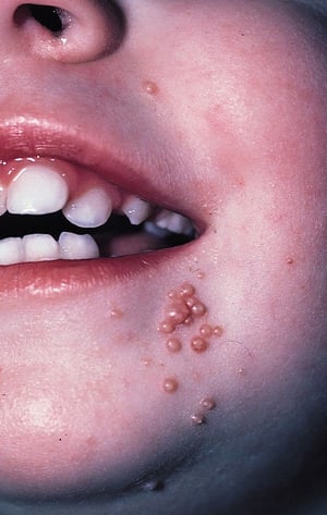
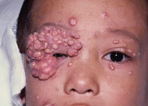
Molluscum contagiosum can appear anywhere on the skin except the palms and soles. Lesions consist of clusters of pink, dome-shaped, smooth, waxy, or pearly and umbilicated papules, usually 2 to 5 mm in diameter, which occur most commonly on the face, trunk, and extremities in children and on the pubis, penis, or vulva in adults. Lesions may grow to 10 to 15 mm in diameter, especially among patients with HIV infection and other immunodeficiencies.
Lesions are usually not pruritic or painful and may be discovered only coincidentally during a physical examination. However, the lesions can become inflamed and itchy as the body fights off the virus.
© Springer Science+Business Media
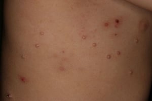
Image courtesy of James G.H. Dinulos, MD.
© Springer Science+Business Media


© Springer Science+Business Media


Image courtesy of James G.H. Dinulos, MD.


© Springer Science+Business Media
Diagnosis of Molluscum Contagiosum
Clinical evaluation
Diagnosis of molluscum contagiosum is based on clinical appearance; skin biopsy or smear of expressed material shows characteristic inclusion bodies but is necessary only when diagnosis is uncertain.
Differential diagnosis includes folliculitis, milia, and warts (for lesions < 2 mm) and juvenile xanthogranuloma and Spitz nevus (for lesions > 2 mm).
Treatment of Molluscum Contagiosum
Physical removal: Curettage, cryosurgery, laser therapy, or electrocautery
Sometimes intralesional injection or photodynamic therapy
Sometimes combination therapies
Most lesions spontaneously regress in 1 to 2 years, but they can remain for 2 to 3 years.
Cantharidin should not be applied to the face or near the eyes because blistering is unpredictable. If cantharidin comes into contact with the cornea, it can cause scarring. Cantharidin should be washed off with soap and water in 6 hours. Fewer than 15 lesions should be treated in one session because infection may occur after application of cantharidin. Parents should be warned about blistering if their children are prescribed this irritant.
Other treatments include intralesional injection (eg, with Candida antigen or occasionally interferon alpha; 2) and photodynamic therapy. Antiviral and immunomodulatory medications have been more successful in patients infected with HIV (3).
Children should not be excluded from school or day care. However, their lesions should be covered to reduce the risk of spread.
Treatment references
1. Shin K, Bae KN, Kim HS, et alJ Am Acad Dermatol pii:S0190-9622(19)32689-1, 2019. doi: 10.1016/j.jaad.2019.08.081
2. Wells A, Saikaly SK, Schoch JJ: Intralesional immunotherapy for molluscum contagiosum: A review. Dermatol Ther 33(6):e14386, 2020. doi: 10.1111/dth.14386
3. Vora RV, Pilani AP, Kota RK: Extensive giant molluscum contagiosum in a HIV positive patient. J Clin Diagn Res 9(11):WD01-2, 2015. doi: 10.7860/JCDR/2015/15107.6797
Key Points
Molluscum contagiosum, caused by a poxvirus, commonly spreads by direct contact (eg, sexual contact, wrestling), fomites, and bath water.
Lesions tend to be asymptomatic clusters of 2- to 5-mm diameter papules that are pink, dome-shaped, smooth, waxy, or pearly and umbilicated.
Diagnose based on clinical appearance.
Treat for cosmetic reasons or prevention of spread.


