


Candidiasis is skin and mucous membrane infection with Candida species, most commonly Candida albicans. Infections can occur anywhere and are most common in skinfolds, digital web spaces, genitals, cuticles, and oral mucosa. Symptoms and signs vary by site. Diagnosis is by clinical appearance and/or potassium hydroxide wet mount of skin scrapings. Treatment is with drying agents and antifungals.
Most candidal infections are of the skin and mucous membranes, but invasive candidiasis is common among patients who are immunosuppressed and can be life threatening.
Systemic candidiasis is discussed in Fungi. Vulvovaginal candidiasis is discussed in Candidal Vaginitis.
Etiology of Mucocutaneous Candidiasis
Potentially pathogenic fungi include dermatophytes and yeast. Candida is a group of about 150 yeast species. C. albicans is responsible for about 70 to 80% of all candidal infections. Other significant species include C. glabrata, C. tropicalis, C. krusei, and C. dubliniensis.
Candida is a ubiquitous yeast that resides harmlessly on skin and mucous membranes until dampness, heat, and impaired local and systemic defenses provide a fertile environment for it to grow.
Risk factors for candidiasis include
Hot weather
Restrictive clothing
Poor hygiene
Infrequent diaper or undergarment changes in children and older adults
Altered flora resulting from antibiotic therapy
Inflammatory diseases (eg, psoriasis) that occur in skinfolds
Immunosuppression resulting from corticosteroids and immunosuppressive medications, pregnancy, diabetes, other endocrinopathies (eg, Cushing disease, hypoadrenalism, hypothyroidism), blood dyscrasias, HIV/AIDS, or T-cell defects
Candidiasis occurs most commonly in intertriginous areas such as the axillae, groin, and gluteal folds (eg, diaper rash), in digital web spaces, on the glans penis, and beneath the breasts. Vulvovaginal candidiasis is common among women. Candidal nail infections and paronychia may develop after improperly done manicures and in kitchen workers and others whose hands are continually exposed to water (see Onychomycosis). In people with obesity, candidal infections may occur beneath the pannus (abdominal fold). Oropharyngeal candidiasis is a common sign of local or systemic immunosuppression.
Chronic mucocutaneous candidiasis typically affects the nails, skin, and oropharynx. Patients have cutaneous anergy to Candida, absent proliferative responses to Candida antigen (but normal proliferative responses to mitogens), and an intact antibody response to Candida and other antigens. They also have impaired T-cell–mediated immunity. Chronic mucocutaneous candidiasis may occur as an autosomal recessive illness associated with hypoparathyroidism and Addison disease (Candida-endocrinopathy syndrome).
Symptoms and Signs of Mucocutaneous Candidiasis
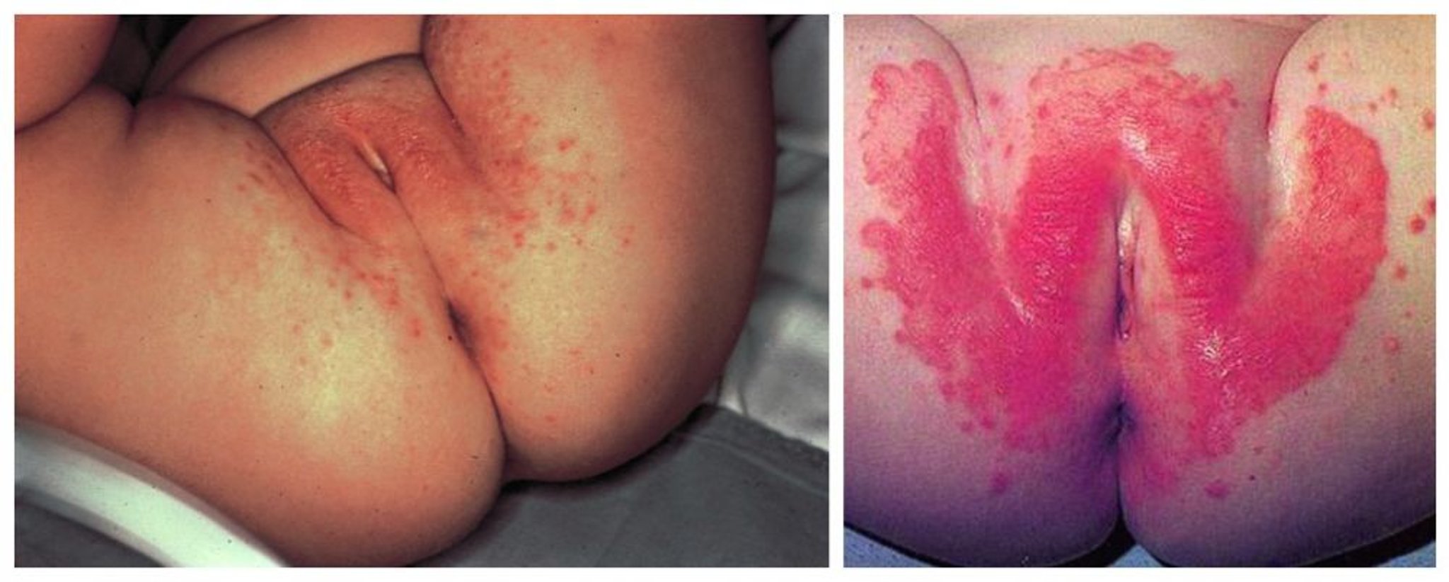
Intertriginous infections manifest as pruritic, well-demarcated, erythematous patches of varying size and shape; erythema may be difficult to detect in darker-skinned patients. Primary patches may have adjacent satellite papules and pustules.
Perianal candidiasis produces white maceration and pruritus ani.

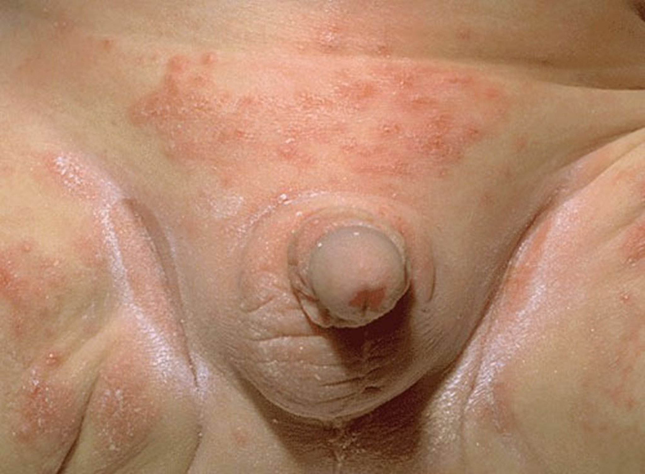
Image provided by Thomas Habif, MD.

© Springer Science+Business Media
Vulvovaginal candidiasis causes pruritus and discharge (see Candidal Vaginitis).


Candidal nail infections can affect the nail plate, edges of the nail, or both. Candidal infection is a frequent cause of chronic paronychia, which manifests as painful red periungual swelling. Subungual infections are characterized by distal separation of one or several fingernails (onycholysis), with white or yellow discoloration of the subungual area.


Oropharyngeal candidiasis causes white plaques on oral mucous membranes that may bleed when scraped (see Interpretation of findings).


Perlèche is candidiasis at the corners of the mouth, which causes cracks and tiny fissures. It may stem from chronic lip licking, thumb sucking, ill-fitting dentures, or other conditions that make the corners of the mouth moist enough that yeast can grow.


Chronic mucocutaneous candidiasis is characterized by red, pustular, crusted, and thickened plaques resembling psoriasis, especially on the nose and forehead, and is invariably associated with chronic oral candidiasis.


Diagnosis of Mucocutaneous Candidiasis
Clinical appearance
Potassium hydroxide wet mounts
Diagnosis of mucocutaneous candidiasis is based on clinical appearance and identification of yeast and pseudohyphae in potassium hydroxide wet mounts of scrapings from a lesion.
Positive culture alone is usually meaningless because Candida is omnipresent.
Treatment of Mucocutaneous Candidiasis
Sometimes drying agents
Topical or oral antifungals
Intertriginous infection is treated with drying agents as needed (eg, Burow solution compresses applied for 15 to 20 minutes for oozing lesions) and topical antifungals (see table Options for Treatment of Superficial Fungal Infections
Candidal diaper rash
Candidal paronychia is treated by protecting the area from wetness and giving topical or oral antifungals. These infections are often resistant to treatment. Thymol 4% in alcohol applied to the affected area 2 times a day is often helpful.
Oral candidiasis
Chronic mucocutaneous candidiasis
Key Points
Candida are normal skin flora that can become infective under certain conditions (eg, excessive moisture, alteration of normal flora, host immunosuppression).
Consider candidiasis with erythematous, scaling, pruritic patches in intertriginous areas and with lesions in the mucous membranes, around the nails, or at the corners of the mouth.
If clinical appearance is not diagnostic, try to identify yeast and pseudohyphae in potassium hydroxide wet mounts of scrapings from a lesion.
Treat most intertriginous candidiasis with a drying agent and a topical antifungal.
Treat most diaper rash with frequent changes of absorbent disposable diapers and an imidazole cream.


